Home

Gentleman win Perfervid cross section of celery under microscope Build on bottle hook

Science | The Crypt School on X: "Some excellent photos taken of a  transverse and longitudinal cross section of a celery. #Y12 #CryptBiology  students had to dissect, stain and view their specimens
Science | The Crypt School on X: "Some excellent photos taken of a transverse and longitudinal cross section of a celery. #Y12 #CryptBiology students had to dissect, stain and view their specimens

wild celery (Apium graveolens), cross section of the stem of celery,  microtome section Stock Photo - Alamy
wild celery (Apium graveolens), cross section of the stem of celery, microtome section Stock Photo - Alamy
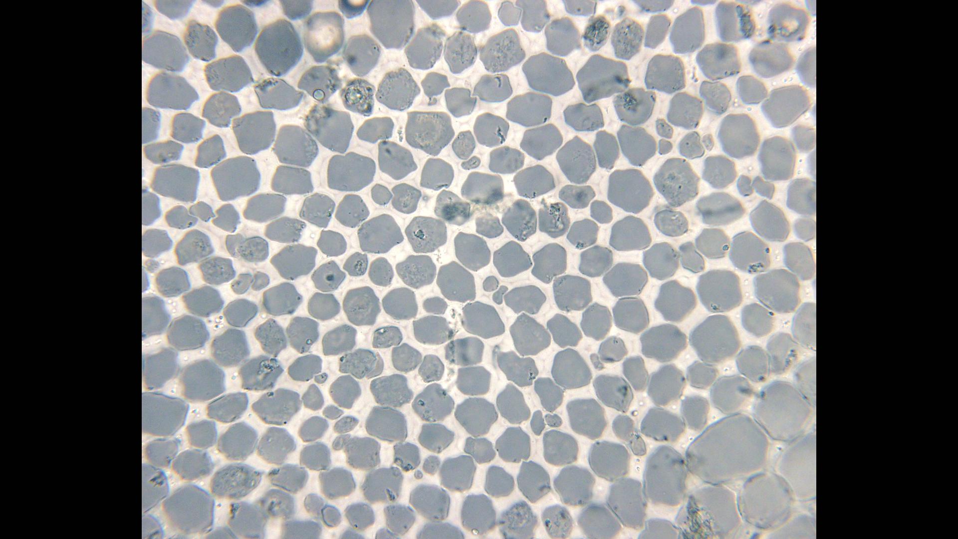
Celery stem, light micrograph - Stock Image - B725/0301 - Science Photo  Library
Celery stem, light micrograph - Stock Image - B725/0301 - Science Photo Library

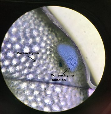
Category: Parenchyma - OSU Plant Structure (BOT313), Winter 2017

Collenchyma tissue - cross section of a celery petiole -100x objective -  UWDC - UW-Madison Libraries
Collenchyma tissue - cross section of a celery petiole -100x objective - UWDC - UW-Madison Libraries

Celery Stripe Cross Section Cut Under Stock Footage Video (100%  Royalty-free) 1017146368 | Shutterstock
Celery Stripe Cross Section Cut Under Stock Footage Video (100% Royalty-free) 1017146368 | Shutterstock

Cross Section Cut Slice of Plant Stem Under the Microscope – Microscopic  View of Plant Cells for Botanic Education Stock Photo - Image of microscopic,  micrograph: 243045478
Cross Section Cut Slice of Plant Stem Under the Microscope – Microscopic View of Plant Cells for Botanic Education Stock Photo - Image of microscopic, micrograph: 243045478

DSC_0237 | Sample: Celery cross section Magnification: 40x S… | Flickr

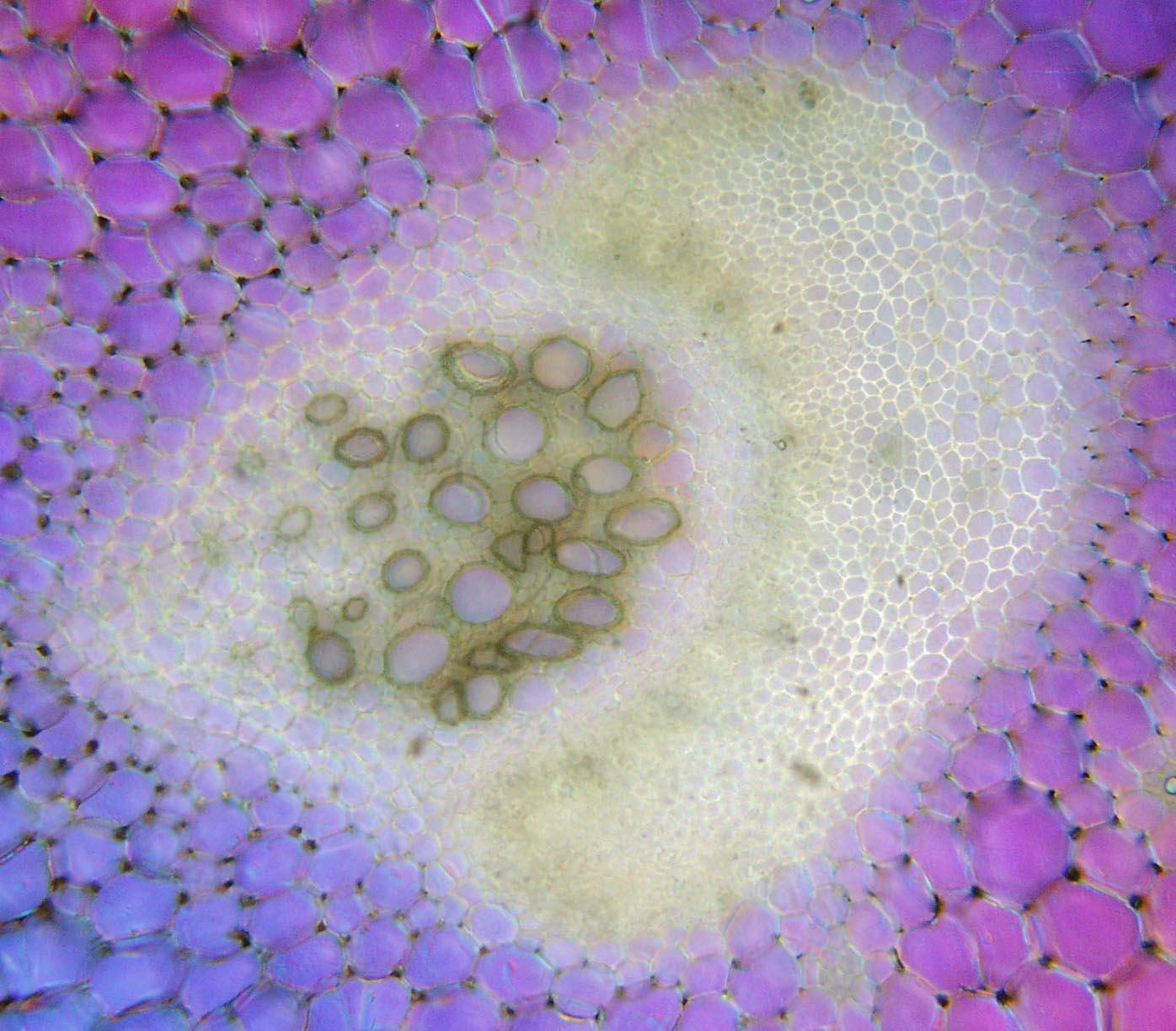
Vascular bundle of cross section of a celery petiole - UWDC - UW-Madison  Libraries
Vascular bundle of cross section of a celery petiole - UWDC - UW-Madison Libraries

Celery Petiole | Cross section showing the Parenchyma and Co… | Flickr

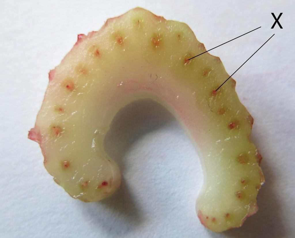
Botany (Grade 5): Celery Stalk Demonstration of the Vascular System – The  Beauty of Play
Botany (Grade 5): Celery Stalk Demonstration of the Vascular System – The Beauty of Play

Gradegorilla Biology

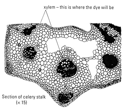
CELERY LAB - Structure and Function of a Plant

The Microscopic Beauty of Plants and Trees by Robert Berdan - The Canadian  Nature Photographer
The Microscopic Beauty of Plants and Trees by Robert Berdan - The Canadian Nature Photographer

Solved D Questions 3 pts List two differences between the | Chegg.com

Observing water moving through plants

Look at the diagram you drew of the celery cross-section under the  microscope. Redraw your diagram and - brainly.com
Look at the diagram you drew of the celery cross-section under the microscope. Redraw your diagram and - brainly.com
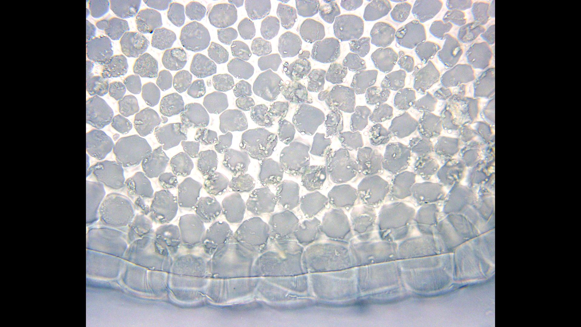
Celery stalk, light micrograph - Stock Image - C007/8467 - Science Photo  Library
Celery stalk, light micrograph - Stock Image - C007/8467 - Science Photo Library

Cross section of a celery petiole - collenchyma tissue inside the epidermis  - 100x objective - UWDC - UW-Madison Libraries
Cross section of a celery petiole - collenchyma tissue inside the epidermis - 100x objective - UWDC - UW-Madison Libraries

Celery Stem Vessel Element Cross Section Stock Photo 1870036333 |  Shutterstock
Celery Stem Vessel Element Cross Section Stock Photo 1870036333 | Shutterstock

General morphology of celery collenchyma (Apium graveolens, eudicot,... |  Download Scientific Diagram
General morphology of celery collenchyma (Apium graveolens, eudicot,... | Download Scientific Diagram
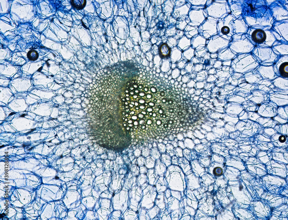
Celery stem with vessel element, cross section, stained with methylene  blue, optical microscpoe. Magnification 160x. Frame width is about 250-300  nm Stock Photo | Adobe Stock
Celery stem with vessel element, cross section, stained with methylene blue, optical microscpoe. Magnification 160x. Frame width is about 250-300 nm Stock Photo | Adobe Stock

CELERY LAB - Structure and Function of a Plant

Solved 1. Parenchyma, collenchyma, epidermis in celery cross | Chegg.com

Celery Lab (Phloem & Xylem) | Biocircuits Outreach

Category: Celery - OSU Plant Structure (BOT313), Winter 2017

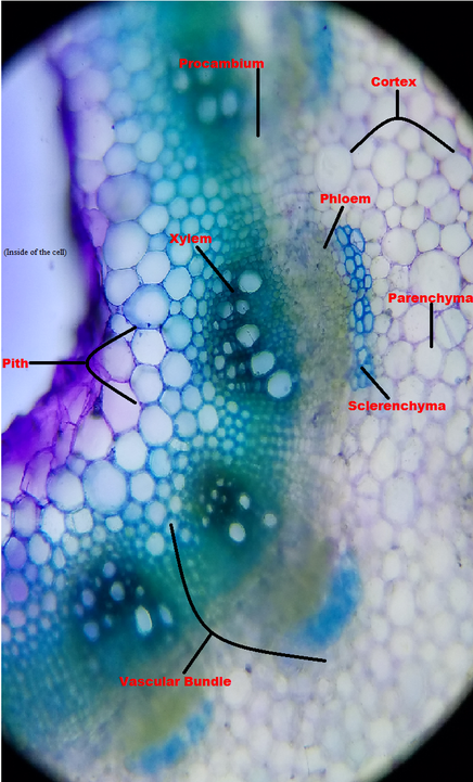
Celery Transport Lab - Celery under the microscope - Tissue identification  Diagram | Quizlet
Celery Transport Lab - Celery under the microscope - Tissue identification Diagram | Quizlet